Extracellular vesicles as diagnostic tools
State-of-the-art fractionation techniques are combined in a customized approach to achieve pure nanovesicle populations at the highest possible yield from various clinical samples (E.g. plasma, urine) or conditioned cell media.
- Differential ultracentrifugation
- Density Gradient Ultracentrifugation
- Size Exclusion Chromatography
- Affinity-based magnetic bead separation
- Fluorescence activated small particle sorting
- Novel experimental techniques
- Commercial kits
Novel detection technologies are developed and benchmarked against state-of-the-art techniques for identification and quantification of extracellular vesicle subpopulations based on protein and/or nucleic acid biomarkers that are predictive of a disease. Methods used for characterization of the biomarkers in the vesicles include:
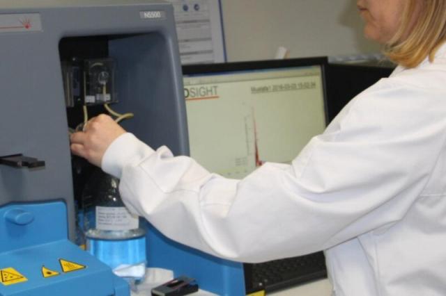
- Fluorescence-based nanoparticle tracking analysis
- Western blot
- (Multiplex) ELISA
- LC-MS-based proteomics platform
- High-resolution flow cytometry for small particle analysis
- Scanning Electron Microscopy
- UV-Vis spectroscopy
- Agilent microarray platform
- Nanodrop, bioanalyzer
DNA mutation detection by hybridization
VITO offers an in-house developed nucleic acid analysis platform that maximizes DNA/RNA hybridization sensitivity while maintaining a high multiplex capability. This is achieved by combining the theory of hybridization with actual wet lab results to allow the optimal design of DNA modified surfaces. The concept is applicable to any hybridization and miniaturized lab-on-chip systems, e.g. for sensitive mutation detection in oncogenes, accurate prediction of antibiotic resistance, and DNA-based monitoring of therapy. In the sample preparation and/or biomarker detection phase nanotechnology is used to enhance detection limits.


